
Fayl:Homeostasis del eritrocito y la hemoglobina.png
Bu prevyuning hajmi: 450 × 600 piksel. Boshqa oʻlchamlari: 180 × 240 piksel | 360 × 480 piksel | 576 × 768 piksel | 768 × 1 024 piksel | 1 361 × 1 814 piksel.
Asl fayl (1 361 × 1 814 piksel, fayl hajmi: 918 KB, MIME tipi: image/png)
| Taʼrif |
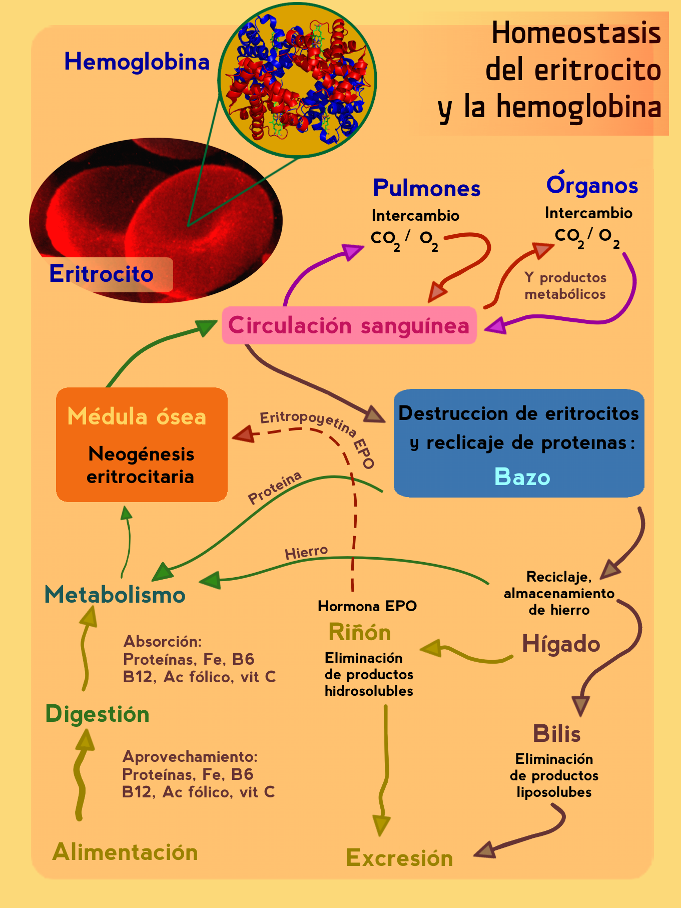
Nueva versión (en español) del gráfico FisiologiaEmoglobina.jpg de Peter Forster. en:Category:Protein images |
| Sanasi | (UTC) |
| Manba | |
| Muallif |
|
| This is a retouched picture, which means that it has been digitally altered from its original version. The original can be viewed here: FisiologiaEmoglobina.jpg:
|
Bu fayl Creative Commons asosida litsenziyalangan Attribution- Share Alike 3.0 Unported litsenziyasi.
- Siz erkinsiz:
- ulashishga – ishlanmani nusxalash, tarqatish va uzatish
- remiks qilishga – ishni moslashtirishga
- Quyidagi shartlar asosida:
- atribut – Siz tegishli litsenziyaga havolani taqdim etishingiz va oʻzgartirishlar kiritilganligini koʻrsatishingiz kerak. Siz buni har qanday oqilona yoʻl bilan qilishingiz mumkin, lekin litsenziar Sizni yoki Sizning foydalanishingizni ma'qullashini taklif qiladigan tarzda emas.
- bir xil ulashish – Agar Siz materialni remiks qilsangiz, oʻzgartirsangiz yoki unga asoslansangiz, oʻz hissalaringizni asl nusxadagi kabi bir xil yoki mos litsenziya ostida tarqatishingiz kerak.
Original upload log
This image is a derivative work of the following images:
- File:FisiologiaEmoglobina.jpg licensed with Cc-by-2.5
- 2006-10-29T06:01:35Z Peter Forster 526x600 (31104 Bytes) {{Information| |Description=Grafica: Medicina: Fisiologia: Ematologia: omeostasi, eritrociti, emoglobina, midollo osseo circolazione sanguigna, milza, fegato, rene, proteine, ferro Fe, eritropoietina EPO, vitamina, B6, B12, C
- 2006-10-28T05:57:48Z Peter Forster 526x600 (31104 Bytes) {{Information| |Description=Grafica: Medicina: Fisiologia: Ematologia: omeostasi, eritrociti, emoglobina, midollo osseo circolazione sanguigna, milza, fegato, rene, proteine, ferro Fe, eritropoietina EPO, vitamina, B6, B12, C
- 2006-10-19T12:22:45Z Peter Forster 526x600 (273771 Bytes) {{Information| |Description=midollo osseo, circolazione sanguina, polmoni, organi, milza, fegato, reni, bile, escrezione, alimentazione, proteine, Fe ferro, vitamina B6, vitamina B12, acido folico, vitamina C, digestione, met
- File:1GZX_Haemoglobin.png licensed with Cc-by-sa-3.0-migrated, Cc-by-sa-3.0-migrated-with-disclaimers, GFDL, GFDL-en, GFDL-self-en
- 2007-06-24T20:54:46Z PatríciaR 1600x1600 (1030152 Bytes) {{Information |Description=By Richard Wheeler ([[:en:User:Zephyris|Zephyris]]) 2007. Created with [[:en:pymol]] from . [[:en:Category:Protein images]] |Source=Originally from [http://en.wikipedia.org en.wikipedia]; descript
- File:SEM_blood_cells.jpg licensed with PD-USGov
- 2006-10-07T20:27:59Z DO11.10 1800x2239 (1398351 Bytes)
- 2006-10-04T03:00:29Z DO11.10 1800x2239 (1012422 Bytes) {{Information |Description=This is a scanning electron microscope image from normal circulating human blood. One can see red blood cells, several white blood cells including lymphocytes, a monocyte, a neutrophil, and many sma
- 2006-10-04T01:09:54Z DO11.10 500x326 (36614 Bytes) {{Information |Description= A three-dimensional ultrastructural image analysis of a T-lymphocyte (right), a platelet (center) and a red blood cell (left), using a Hitachi S-570 scanning electron microscope (SEM) equipped with
Uploaded with derivativeFX
Fayl tarixi
Faylning biror paytdagi holatini koʻrish uchun tegishli sana/vaqtga bosingiz.
| Sana/Vaqt | Miniatura | Oʻlchamlari | Foydalanuvchi | Izoh | |
|---|---|---|---|---|---|
| joriy | 01:57, 6-fevral 2010 | | 1 361 × 1 814 (918 KB) | Neotex555 | cambioa version en PNG |
| 01:45, 6-fevral 2010 |  | 1 361 × 1 814 (367 KB) | Neotex555 | {{Information |Description=Nueva versión del gráfico FisiologiaEmoglobina.jpg de Peter Forster. Pedido por [http://es.wikipedia.org/wiki/Usuario:Rjgalindo] en:Category:Protein images This is a scanning electron microscope image from normal circulat |
Fayllarga ishoratlar
Bu faylga bogʻlangan sahifalar yoʻq.
Faylning global foydalanilishi
Ushbu fayl quyidagi vikilarda ishlatilyapti:
- ca.wikipedia.org loyihasida foydalanilishi
- es.wikipedia.org loyihasida foydalanilishi
- kn.wikipedia.org loyihasida foydalanilishi